Dogs and cats with skin conditions or lesions can be a challenge to diagnose, especially when the condition is chronic. Investigations can include screening for systemic illness, assessing for food sensitivities and atopy, but useful information can also be gained from examining superficial skin preparations under the microscope. Here, we will look at some different sample types and some of the abnormalities they can identify.
Skin Scrapes
When performing a skin scrape, clipping the long hair over the lesion may help the sampling process, but try and leave crusts/ scales behind as these will be part of the sample. Gently squeezing and rolling the skin before scraping may help to surface some deeper pathogens (such as Demodex sp). The skin scrape should ideally go down to capillary ooze for adequate depth to find Sarcoptes sp. if present. The material obtained from the scrape can then be transferred to a slide – a drop of liquid paraffin can help to anchor the material on the slide. The material can then be thinned out on the slide and a coverslip (ideally large) applied. Very thick areas may be of limited value as it is difficult to see the elements within them.
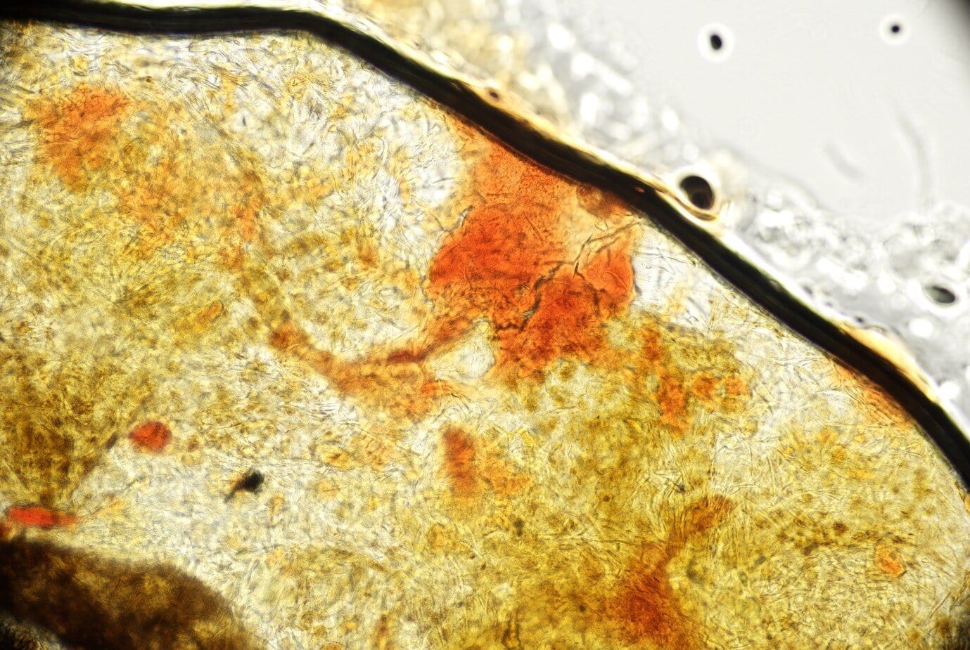
Figure 1. Skin scrape, x 10 objective, showing red blood cells indicating adequate depth of preparation.
Scan over the slide on x 10, increasing to x 20 when areas of interest are identified. Skin scrapes are classically used to identify mites. Demodex sp. may be easier to find than Sarcoptes sp. as they are often present in higher numbers.
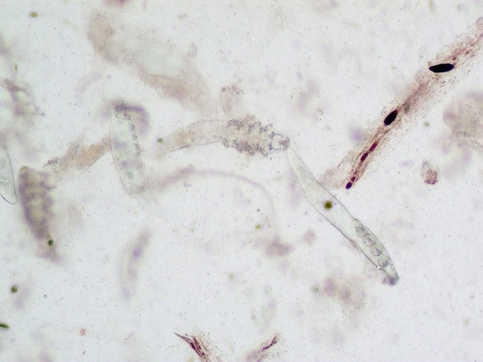
Figure 2. Elongate Demodex sp. mites with long tails.
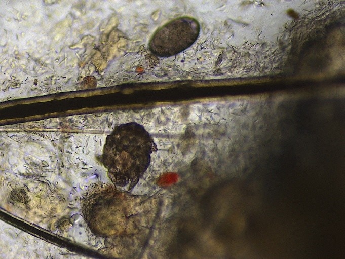
Figure 3: Sarcoptes sp. mite (bottom) and Sarcoptes sp. egg (top). Rounded mites with visible legs. Note the size of the hair between the two – hairs can be used to give a sense of scale.
Different species of Demodex sp. have different length tails – for example, Demodex cati have long tails and Demodex gatoi have short tails. The latter can cause clinical signs with a lower number of organisms and may be contagious between cats.
Sarcoptes sp. may be difficult to find on skin scrapes as low numbers of organisms can cause severe disease – submitting multiple scrapes may be of value, and if results are negative but a clinical suspicion remains, consider serology as an alternative.
Skin scrapes and hair plucks may both yield evidence of dermatophyte infections. These can be seen as macroconidia (club or cigar shaped elements) with internal segmentation. These should ideally be followed up with culture or PCR to confirm and speciate as some environmental contaminants may appear similar. Microsporidia can be found within or bursting out of hair shafts.

Figure 4: Macroconidium, likely Trichophyton sp. x 20
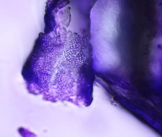
Figure 5: Microconidia, likely Trichophyton sp. x 100
Tape Strips
Applying a stretch of clear tape (not micropore!) to superficial skin lesions helps lift off the most superficial layer and, once stained with a rapid stain, can be useful in looking for superficial inflammation and bacterial or yeast organisms. Some abnormalities of the superficial keratinized skin cells can also be detected.
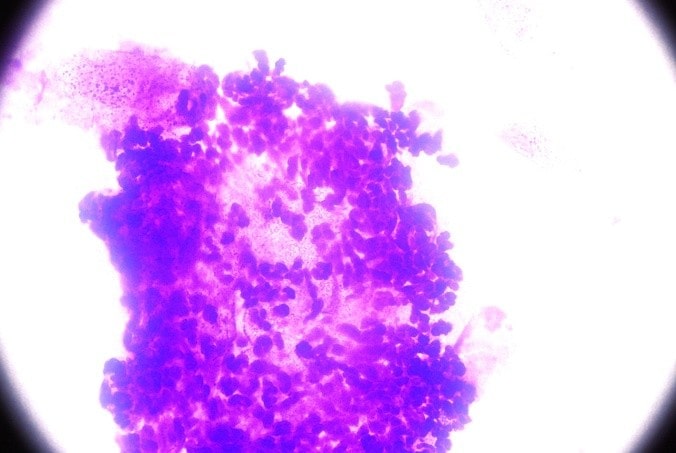
Figure 6: Neutrophilic inflammation (pyoderma) in a tape strip sample. 100x
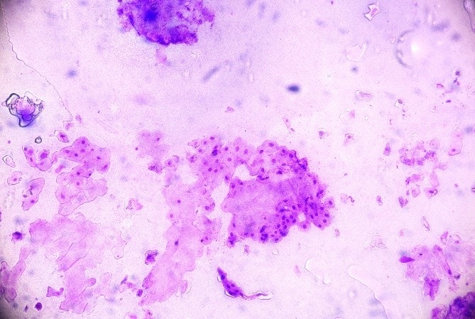
Figure 7: Nucleated squamous epithelial cells (parakeratosis) in a tape strip. This may be associated with increased skin cell turnover with a few different conditions including parasitic, immune mediated disease and some dietary deficiencies. 20x.
Hair Plucks
Hair plucks are gathered by grasping groups of hairs and pulling them out by the roots. The shape of the hair roots can be assessed for shape and appearance to determine if these are resting (telogen) or growing (anagen) hairs. Obtaining all telogen hairs, for example, may mean that an underlying endocrinopathy could not be ruled out as a cause of hair thinning/ alopecia. Hairs can also be inspected for dermatophytes. The distribution of pigment within the hairs may be spotty/ splodged in colour dilute (e.g. ‘blue’ coat colour) which can be associated with colour dilute alopecia
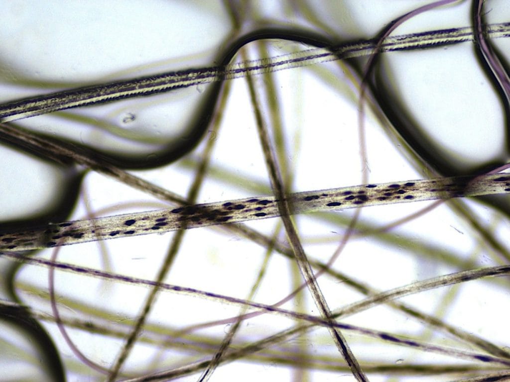
Figure 8: The top horizontally orientated cell has normal pigment distribution. The splotchy pigment in the horizontal hair below this is typical of a colour dilute individual.
Hair tips can also be assessed for fraying associated with the trauma of a pruritic individual chewing the area.
Although this is by no means a complete appraisal of hair/ skin microscopy, this illustrates some of the useful findings from these superficial samples.

