Digestive disorders are a frequent reason that pets are presented to veterinary practices, with the main complaints being, vomiting, anorexia, flatulence, weight loss and diarrhoea. Diarrhoea is described as an increase in the frequency and volume of faeces, there may be a mixture of blood and/or mucus. If the diarrhoea lasts less than 3 weeks, it is classified as acute. If the problem persists for longer than 3 weeks, it is a chronic or chronic-recurrent digestive disorder. In most cases, acute diarrhoea is self-limiting and can be successfully treated with diet and/or fluid substitution. Often there are no apparent reasons for the diarrhoea. In patients with persistent digestive disorders, however, a detailed diagnostics must be carried out. Clarity must be obtained on understanding whether or not the cause is one of primary gastrointestinal (localised in the gastrointestinal tract) or extra gastrointestinal origin.
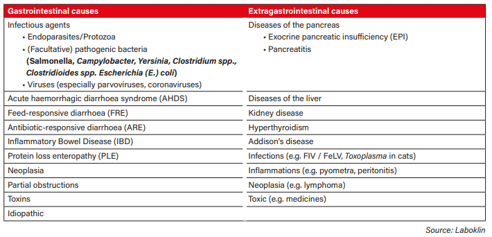
Table 1: Possible causes of indigestion
Table 1 provides an overview of possible gastrointestinal and extra gastrointestinal causes.
The initial history and clinical examination of the patient precedes any laboratory diagnosis. Apart from the standard history such as breed, age, vaccination status, deworming, current medication, previous treatments, etc., the information on the duration of symptoms, faecal consistency and frequency of defecation. In this way, it may be possible to narrow down whether the problem is originating in the small intestine or large intestine (or a mixed form) (Table 2). The information from the Initial history and the clinical examination of the patient together form the basis of which laboratory diagnostic tests are selected.
The choice of laboratory diagnostic tests depends, on whether acute or chronic diarrhoea is present and which previous examinations have been carried out.
The initial blood and faecal examinations will provide information on further decisions and what follow-up examinations (imaging or elimination diets) should be done.
BASIC EXAMINATIONS
Basic examinations include:
Faeces
•Parasitological examination (endoparasites)
•Examination for facultative intestinal pathogens
•Examination for viral pathogens
Blood
•Haematology
•Blood chemistry
If a patient with diarrhoea comes to the veterinary practice for the first visit, a parasitological examination should be carried out. An infestation of intestinal parasites such as roundworms, whipworms, or hookworms can lead to gastrointestinal disorders, as can an infestation protozoal infections such as giardia or coccidia. The examination is carried out microscopically (flotation/sedimentation) or, in the case of giardia, also by means of an ELISA (immunological method). A 3-day pooled faecal sample increases the probability of detecting intermittently excreted pathogens. In cats, a possible infection with Tritrichomonas foetus should be considered, which can be identified by means of a PCR examination. In cases of acute diarrhoea, especially in young animals or in connection with a disturbed general condition (e.g., fever), various viral pathogens (parvoviruses, circoviruses, coronaviruses) can be the cause. These can partly be examined by ELISA and/or PCR. A bacteriological examination of the faecal sample may be considered to identify an infection of (facultative) intestinal pathogens. These include Salmonella spp, Yersinia spp, Campylobacter spp, E. coli, and gas-forming bacteria such as Clostridium spp. and Clostridioides sp. Clostridium perfringens and Clostridioides difficile are capable of forming various enterotoxins. It makes sense to test dogs and cats fed according to the BARF concept for Salmonella, Yersinia, Campylobacter, and Listeria. In addition to the bacteriological examination, the mycological examination can also give indications of a dysbiosis through an increased detection of yeast fungi.
With the help of blood tests (Full Blood Count (FBC) and chemistry), it is possible to find out whether there is a systemic disease associated with gastrointestinal complaints.
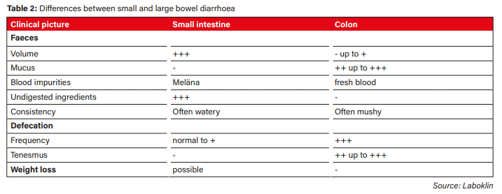
Table 2: Differences between small and large bowel diarrhoea
Blood tests ( Full blood count and chemistry), will assist in identifying whether or not there is a systemic disease,(such as liver or kidney disease) dehydration or a parasitic infection present. Indications of an inflammatory process can also be detected. It is also useful to determine the total protein and albumin, for example, to detect protein losing enteropathy or other diseases that are also associated with protein loss.
FURTHER INVESTIGATIONS
If the first examinations have not yet led to a diagnosis, further differential diagnoses must be explored. Further examinations include:
Faeces
•Presence of maldigestion / malabsorption
– microscopic food utilisation
– canine pancreatic elastase (dog)
– Bile acids (chologenic diarrhoea)
•Presence of an inflammatory event
– Calprotectin
•Presence of protein loss
– alpha-1-antitrypsin
•Presence of dysbiosis
– Dysbiosis analysis
Blood
•Pancreas
– TLI, vitamin B12, folic acid
– PLI
•Hyperthyroidism
– T4 (cat)
•Addison’s disease
– Basal cortisol, Na/K ratio
•Infections (Cat)
– Antibodies (FIV, Toxoplasma), FeLV antigen
Microscopic food utilisation is used to assess undigested food components such as fat, muscle fibres, and starch by means of a special stain and subsequent microscopy of the faeces. This examination is not specific to a particular clinical picture, but can give indications of reduced digestive and absorptive capacity (maldigestion or malabsorption). In the case of weight loss with vomiting and diarrhoea, the canine pancreatic elastase can be used to check the function of the exocrine pancreas. This parameter can be used as a screening for exocrine pancreatic insufficiency. It is an enzyme that is secreted by the pancreas and is not broken down in the intestine. A high value confirms sufficient function of the exocrine pancreas. In the case of an EPI, the values are low – but these can also be temporarily low in dogs with diarrhoea due to a thinning effect in the faeces and in healthy dogs. Therefore, in these cases, the TLI (trypsin-like immunoreactivity) concentration in the blood should be determined.
Calprotectin is a biomarker that can indicate inflammatory processes in the intestine. It is a protein that is mainly formed in neutrophil granulocytes. If there is an inflammatory stimulus, more granulocytes penetrate the intestinal wall via diffusion and thus increase the calprotectin concentration in the faeces. The determination of alpha-1-antitrypsin can be carried out if protein loss via the intestine is suspected. Alpha-1-antitrypsin is a protease inhibitor that is similar in size to albumin and is lost via the intestine in approximately the same way, for example if there is increased permeability of the intestinal wall. In contrast to albumin, it is insensitive to bacterial proteolytic degradation in the intestine and is thus excreted unchanged in the faeces. Increased values indicate an enteric protein loss. A dysbiosis analysis can help clarify whether the intestinal microbiota is imbalanced. If the composition of the intestinal flora is disturbed or if there is a shift among the commensal pathogens, this is called dysbiosis. Increasingly, various clinical pictures, such as chronic diarrhoea, food intolerances, chronic enteropathy or metabolic complaints, are associated with dysbiosis. Only a very small proportion (< 1 %) of bacteria can be detected by cultural cultivation, although molecular biology-based dysbiosis analysis (qPCR) can also detect pathogens, regardless of their cultivation conditions (especially anaerobes).
Other differential diagnoses can be specifically identified with certain parameters from the blood. Diseases of the pancreas should be diagnosed by examining the TLI concentration, the PLI concentration (pancreatic lipase immunoreactivity), vitamin B12 (cobalamin), and folic acid. The determination of TLI is a specific test for the detection of exocrine pancreatic insufficiency (TLI concentration decreased). The PLI value is a biomarker for the diagnosis of pancreatitis. An increase in the PLI concentration in the blood is indicative. Endocrinopathies, e.g. hyperthyroidism in cats, can be clarified by determining the total thyroxine (T4). In Addison’s disease, electrolyte shifts (sodium/potassium quotient) and a lowered basal cortisol level are visible (note: there are also atypical forms without electrolyte shifts).
Infections with feline immunodeficiency virus (FIV) and feline leukaemia virus lead to immunosuppression in the cat and thus predispose to infections that can affect the gastrointestinal tract. The detection of FeLV antigen and FIV antibodies is carried out via a ELISA or the detection of the provirus via PCR. Imaging examinations of the abdomen, histopathological examinations of tissue samples, and the elimination diet are further important diagnostic methods for clarifying chronic diarrhoea. Histological examination can be helpful in the determination of a suspected tumour. In addition, typical changes can give indications of a protein loss enteropathy as well as general information about an inflammatory process in the intestine (e.g., feed intolerance, IBD). An elimination diet is a non-invasive way to clarify food-responsive diarrhoea. Strict adherence to the diet is of great importance and should be realistically assessed depending on the owner’s compliance and the animal’s environment (e.g., doubtful for a family dog in a family with small children from whom the dog picks up food scraps).
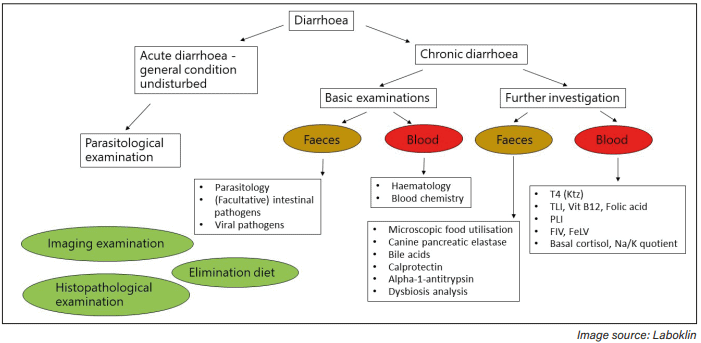
Fig. 3: Diagnostic work-up diarrhoea
SUMMARY
Figure 3 gives an overview of a possible diagnostic work-up in patients with diarrhoea.
Dr Corinna Hader
FURTHER READING
- Dahlem D, Burgener I. Chronische Diarrhoe. kleintier konkret 2015; 18(03): 28-39. doi: 10.1055/s-0035-1550096.
- Dorn D, Mangelsdorf S.Pankreatitis beim Hund. kleintier konkret 2018; 21(01): 20-31 doi: 10.1055/s-0043-124118.
- Ewald N, Rödler F, Heilmann RM. Chronische Enteropathien bei der Katze – diagnostische und therapeutische Aspekte. Tierarztl Prax Ausg
- K Kleintiere Heimtiere 2021; 49(05): 363-376.doi: 10.1055/a-1584-9705