
When you take a cytology samples from a mass, do you fear you will get a non-diagnostic result?
This month, we are going through some tips about how to obtain good quality, diagnostic cytology samples and possibly avoid the much-feared interpretation.
- The first hack is about speediness. Make sure that when you are taking your samples, you have everything you need at hand, such as few needles, a syringe, multiple glass slides and a pencil. We recommend to wite on the frosted end of every smear where the sample was taken from by using a pencil. This because the pencil lead does not dissolve during the staining process, as pen and marker ink do.
Then, you have to assess the type of lesion you are dealing with, in order to select the most appropriate sampling techniques.
- For small, soft or vascular lesions, the fine needle biopsy (FNB) is the preferred method. Hold the lesion between your fingers, and using your free hand, stick the needle (22-21 gauge) inside the lesion. Rotate the needle within the lump, move it up and down and back and forth, to collect as many cells as possible. In this short video, you might able to better appreciate how to perform the procedure.
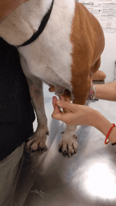 (click on the picture to view a video)
At this point, you can remove the needle, attach it to a syringe filled with air and aspirate the collected material onto a clear slide.
FNB technique is easy to perform, minimises tissue and patient trauma and, most importantly, it reduces blood contamination to a minimum, as no suction is applied. A possible downside might be the relatively low numbers of cells entering the needle only through capillarity action, but this caveat can be overcome by taking multiple aspirates from the same lesion.
- If the mass is harder, fine needle aspiration (FNA) is the way to go. In this case, you must pierce the mass with a 22-21-gauge needle attached to a syringe. Then withdraw the plunger creating vacuum and without exiting the lesion, redirect the needle along several different tangents. Before exiting the mass, release the plunger to halt the vacuum. At this point, you detach the needle from the syringe, fill it with air, re attach the needle and squirt the sampled material onto a slide. This video will help you to master the technique.
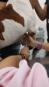 (click on the picture to view a video)
FNA technique certainly will retrieve more cells, especially from hard tissue such as bone, however there is higher risk of having reduced cell preservation and haemodiluted samples.
- If you are suspicious for scabies or dermatophytosis, the most suitable sampling method is skin scraping performed with a surgical blade. In particular, for Demodex, it is recommended to squeeze the skin between your fingers while scratching it with the blade, possibly inducing a little bleeding. The purpose of it is to pull out Demodex mites that tend to nest into the deepest parts of hair follicles.
- For corneal lesions, vaginal cavity, ear canal or fistulae, swabbing is the way to go. If you are sampling a very dry surface, it might be helpful to moisten this with some sterile saline beforehand.
- Finally, an impression smear is the preferred method for ulcerated lesions or biopsy samples. Remember do not disinfect the ulcerated lesion prior to sampling, as you want to sample everything (including the junk!) (see Picture 1). On the contrary, if you are dealing with a biopsy, dab it with a clean gauze to dry away as much blood as possible and then press it multiple times onto the slide (see Picture 2).
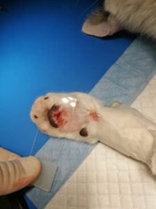
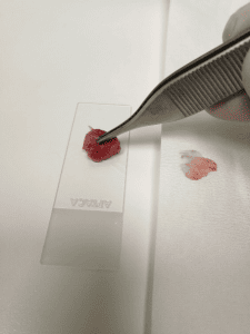
Picture 1: Impression smear from an ulcerated lesion Picture 2: Impression smear of a biopsy