PART I: BEVA CONGRESS, SEPTEMBER 2023, BIRMINGHAM, UK
HOPSTER-IVERSEN: WEIGHT LOSS – CAN WE ONLY PRESCRIBE STEROIDS?
The author addressed the topic of chronic weight loss in adult horses and discussed the subject of inflammatory bowl disease (IBD) in particular. This is characterised by the infiltration of inflammatory cells into the mucosa or submucosa of the GI tract. IBD must be diagnosed by diagnosis of exclusion and the following criteria must be fulfilled: thickened intestinal walls (sonography), abnormal absorption test (caution: large intestine), hypoproteinaemia and hypoalbuminaemia and histological evidence of infiltration of inflammatory cells (lymphocytes, eosinophil granulocytes, etc.). The aetiology has not yet been clarified. An immune-mediated reaction to an as yet unidentified antigen is suspected. One therapy is to avoid the suspected antigen. An elimination diet and possibly endoparasite control are also recommended. Systemic dexamethasone may be tried initially, followed by prednisolone (sometimes lifelong). While the prognosis is moderate, if treatment is started early, horses with persistent malabsorption have a limited chance of recovery.
HUGHES: TRANSFAUNATION – IS FAECES THE CURE?
Hughes addressed the topic of faecal microbiota transplantation in horses. Indications are diarrhoea, enteritis and colitis – even in foals older than 24 hours. Donor horses should be healthy, show received no medication in recent weeks and have a negative faecal screening for pathogens. The preparation of faeces has been described in literature. Adult horses receive 2 – 7 litres, foals 400 – 600 millilitres.
SULLIVAN: COLITIS IN DONKEYS
Not only did the author point out donkey-specific deviations in behaviour, but also in laboratory diagnostic parameters. Donkeys can be triggered by all forms of stressors. Also in donkeys, transfaunation yielded good results. Only a healthy donkey can be considered as a donor. Probiotics used in horses should not be used, as donkeys have a different microbiome than horses.
SOUTH: HEPATOTROPIC VIRUSES – WHERE ARE WE TODAY?
Liver diseases caused by hepatotropic viruses were discussed in detail. Theiler’s disease is a liver disease that usually occurs acutely and quickly becomes life-threatening.
It is caused by an infection with the equine Parvovirus (EqPV-H), which in most cases is is transferred to horses when equine biologics are used. If this is not the case, other transmission routes are discussed (blood-sucking vectors?). T. Divers isolated equine parvovirus as an aetiological agent a few years ago, but Theiler’s disease has been described for much longer. In early stages, the increase in γ-GT is not alarmingly high. According to one study, the seroprevalence is around 30 %, and 9 % show a positive PCR test result. Infection with equine Hepacivirus (EqHV) is usually subclinical. However, clinical diseases have been triggered experimentally.
Rossdales Laboratories found 2.6 % Hepacivirus-positive results (serology and/or PCR) among 227 sample submissions from healthy horses, while the prevalence for equine Parvovirus was < 1 %. This raises the question of the significance of these pathogens as a cause of hepatopathies. It becomes clear that a single positive detection does not allow an aetiologically clarified diagnosis.
VAN ERCK-WESTERGREN: ELEVATED LIVER ENZYMES IN SPORT HORSES – SHOULD WE BE WORRIED?
The author discussed the significance of elevated liver enzymes, especially γ-GT, in sport horses. The “γ-GT syndrome”: γ-GT is moderately elevated after intensive, cumulative training sessions. Oxidative stress is suspected as the cause. With increased O2 consumption, 98 % of the oxygen is used to provide energy, while 2 % remains in the body as highly reactive oxidative stress metabolites (ROS). This accumulation of ROS can be interpreted as training intolerance.
CLAES: POSSIBLE USE OF REPRODUCTIVE ENDOCRINOLOGY IN THE DIAGNOSTICS BY MARES
The author presented the following laboratory approach to the diagnosis of granulosa cell tumours (GCT): Testosterone determination: 48 % sensitivity (adrenal or ovarian origin, increased in pregnant mares, daily variations). Inhibin B determination: 80 % sensitivity (cyclical changes, increased during pregnancy). Inhibin and testosterone determination: 84 % sensitivity. Anti-Müllerian hormone (AMH) determination showed a sensitivity of 98 %, and was recommended for CCT diagnostics! In males, AMH drops to gelding level approx. one week after castration (t1/2: 1.5 days).
RENDLE: IS ERTUGLIFLOZIN A MIRACLE DRUG?
The author summarised Sundra’s work on ertugliflozin once again. Her results were supported by further studies and have been confirmed: Ertugliflozin appears to be a useful drug for treating EMS and laminitis, and will take EMS therapy to a new level. It was not only the weight loss, the drop in insulin concentration and the significant improvement in laminitis symptoms within a short period of time that were impressive, but also the owner satisfaction in horses that were already being considered for euthanasia was extremely positive. However, it should be noted that some horses, which are returned to normal use, already have structural changes in the hoof due to a long history of laminitis. The long-term development of these horses must be monitored.
SULLIVAN: ENDOCRINOPATHIES IN DONKEYS
The author mainly presented essentials about the donkey metabolic syndrome (AMS). The owner’s assessment of the animal’s nutritional status is severely impaired. 26 % of donkeys in the UK are overweight, almost 9 % are obese and this problem is increasing. The diagnosis of AMS (asinine metabolic syndrome) is the same as for horses, but with adjusted reference values. The Karo light syrup® test has not yet been validated for donkeys. According to the author, the cut-off for insulin should be around 50 μU/ml. The treatment includes: diet, exercise (even donkeys can be exercised!), 2 to 3 kg roughage/day (in winter: 50 % straw, in summer: 75 % straw) and appropriate feed supplements (balancers).
HUGHES: ROTAVIRUS B – THE “NEW KID ON THE BLOCK”
A new Rotavirus B has led to outbreaks of bloody diarrhoea in young foals at several stud farms in Kentucky. The source of this new equine Rotavirus group B (ERVB) was ruminants. PCR testing for the known ERVA did not detect the new virus. The foals were 2 – 7 days old, the diarrhoea lasted for 3 – 4 days and the morbidity rate was up to 100 %. Previous vaccination of the mares with an inactivated ERVA vaccine did not protect the foals against ERVB disease. So far, no commercial test for ERVB is available, so it remains unclear where else the virus may be present. A certain zoonotic potential of ERVB cannot be ruled out.
PART II: ANNUAL CONVENTION OF THE AMERICAN ASSOCIATION OF EQUINE PRACTITIONERS (AAEP), DECEMBER 2023, SAN DIEGO, CA, USA

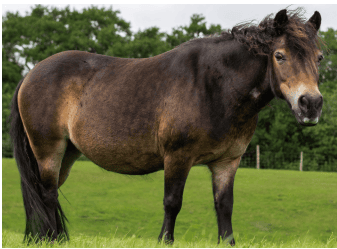
Fig. 2: Overweight pony
VARIOUS IMPORTANT PUBLICATIONS FROM THE PAST YEAR WERE PRESENTED AT THE “KESTER NEWS HOUR”:
Boger et al. investigated the effect of a single intraarticular (i. a.) triamcinolone acetate (TA) injection on blood glucose and insulin concentrations in horses to evaluate the risk of laminitis following cortisone administration. For the study, insulin and glucose concentrations were determined basally in 10 healthy horses and after an i.a. TA injection (measurements after 4, 6, 8, 24, 48 and 72 h). Moderate increases compared to the basal values were observed up to 48 h after TA injection. The study showed that an i.a. cortisone injection can influence blood glucose and insulin levels up to 48 h after injection in healthy horses. In horses with suspected insulin dysregulation (ID), this should be taken into account prior to a TA injection. It is recommended that they are tested for ID – e.g. using the Karo light syrup® test or oral glucose test – in order to better assess the risk of laminitis.
Dr Noah Cohen (Texas School of Veterinary Medicine) presented a new research project: The development of an mRNA vaccine against Rhodococcus hoagii (formerly R. equi) infections in foals with the aim of improving their immune response.
In another study, Stratico et al. investigated owner satisfaction following ovariectomy in 11 mares (unilateral n = 5, bilateral n = 6) with behavioural abnormalities (e. g. unrideability, increased sensitivity on both flanks, etc.). After the surgery, the majority of owners (91 %, 10/11) reported a significant improvement in behaviour (reduction in severity or complete disappearance of the abnormalities).
SCIENTIFIC LECTURES
Neurological and neuromuscular diseases in horses were this year increasingly discussed. Amy L. Johnson (University of Pennsylvania) cited cervical vertebrae stenotic myelopathy (CVSM or Wobbler syndrome) and equine neuroaxonal dystrophy (eNAD)/equine degenerative myeloencephalopathy (EDM) as the causes of the most common non-infectious spinal cord diseases in horses in the USA with spinal ataxia. eNAD and the advanced form of this disease (EDM) are neurodegenerative disorders with a genetic predisposition that are associated with a vitamin E deficiency within the first year of life. In addition to performance deficits, ataxia and proprioceptive deficits, horses may show behavioural and/or temperamental changes. These vary from sudden, unpredictable explosive behaviour (aggression, bucking, unrideability, etc.) to lethargy or sluggishness. An ante mortem diagnosis is difficult and therefore a diagnosis of exclusion is required: a CSF examination usually shows no evidence of an inflammatory-infectious process. CVSM should be excluded by means of X-ray examinations. The examination of the biomarker pNF-H (phosphorylated neurofilament heavy chain protein) from CSF and serum can be useful to support the diagnosis of eNAD/EDM ante mortem. This protein indicates neuroaxonal degeneration and the subsequent release of structural proteins (neurofilaments). In addition, serum vitamin E levels are often marginal to low and are cited as a risk factor for eNAD/EDM. Since only histopathological changes in the brain stem and spinal cord allow a clear diagnosis post mortem, the diagnosis of eNAD/EDM tends to be underdiagnosed. Amy L. Johnson cites this disease as the most common neurological diagnosis post mortem.
Equine motor neuron disease (EMND) – an acquired, neurodegenerative disease of the somatic lower motor neurones that innervate the skeletal muscles – is also a vitamin E-associated muscle disease of middle-aged horses.
The horses show no ataxia, but weakness and generalised muscle atrophy. Diagnosis is based on a biopsy of the sacrocaudalis dorsalis medialis muscle and a determination of the vitamin E concentration in the serum. It must be taken into account that normal vitamin E concentrations cannot rule out previous deficiencies.
Vitamin E responsive myopathy (VEM), a subtype of EMND, is a mostly reversible disease that is associated with muscle atrophy and weakness, but does not cause damage to the motor nerves. However, the vitamin E determination can show normal to low serum values. A distinction between EMND and VEM can only be made on the basis of a biopsy of the sacrocaudalis dorsalis medialis muscle. While EMND patients often do not respond or only respond to a limited extent to treatment with vitamin E or show permanent muscle atrophy, patients with VEM respond very well to treatment with vitamin E – to the point of complete recovery.
The most common infectious cause of spinal cord disease in horses in the USA is equine protozoal myeloencephalitis (EPM), caused by a protozoal infection (Sarcocystis neurona, rarely also Neospora hughesi). Symptoms include lameness, proprioceptive ataxia, paresis and asymmetric muscle atrophy. Due to the very high seroprevalence (up to 83 % depending on the region), a positive antibody titre should not be used as basis for a diagnosis. Amy L. Johnson therefore recommends ante mortem exclusion diagnostics based on the examination of a paired serum sample, the antibody titre ratio of serum/cerebrospinal fluid, PCR examination of cerebrospinal fluid and diagnostic therapy. EPM should definitely be considered as a differential diagnosis in imported animals from the USA in the case of unclear orthopaedic and neuromuscular diseases.
The diagnosis of polysaccharide storage myopathy type 2 (PSSM2) and myofibrillar myopathy (MFM) is made by biopsy of the semimebranosus or gluteus medius muscle. Stephanie Valberg (Kentucky Equine Research) describes the so-called “Percutaneous Needle Biopsy Technique”. This and other biopsy techniques are described clearly and simply on the website of the Valberg Neuromuscular Diagnostic Laboratory.
According to Amy L. Johnson, Borrelia burgdorferi plays a minor role as a cause of neurological disease in horses. However, she reported regularly occurring cases of involvement of this pathogen in nuchal bursitis. The diagnosis is made via a positive PCR test from bursal fluid. In the lecturer’s experience, these horses are often also highly positive for OspA antibodies in the Western blot, which are actually more likely to be associated with vaccination against the pathogen.
REPRODUCTION
Progesterone, 5ɑ-dihydro-progesterone (DHP) and the significance for the luteo-placental shift in pregnancy:
Alan Conley (University of California, Davis) spoke about the importance of determining these hormones during pregnancy using liquid chromatography with tandem mass spectrometry (LC-MS/MS). In particular, the course of progesterone and DHP and the ratio of these two hormones in early pregnancy can be relevant for practitioners in order to determine the so-called luteo-placental shift – i. e. the time at which the placenta takes over progestogen synthesis. Luteal progesterone synthesis decreases between the 70th and 100th day of pregnancy. Instead, the chorioallantoic membrane produces DHP until birth. A distinction between these two hormones is only possible using LC-MS/MS. In cyclic or early pregnant mares, the DHP/progesterone ratio is < 1, whereas it is > 1 after the luteo-placental shift (around the 100th to 120th day). In mares with suspected luteal insufficiency (progesterone < 4 ng/ml), determining the time of the luteo-placental shift may be relevant in order to estimate the treatment period of Altrenogest supplementation. Due to the androgenic properties of Altrenogest, with possible effects on the long-term fertility of the mare and also on newborn fillies, it should not be given for an unnecessarily long period. The determination of the DHP/progesterone ratio can therefore be helpful in making a therapy decision. Conventional immunoassays can only determine total progesterone due to cross-reactivity. Determination of all relevant progestogens using LC-MS/MS can therefore be advantageous in cases of suspected luteal insufficiency. Furthermore, if the DHP concentration is sufficient, it can be assumed that supplementation with Altrenogest can be stopped without risk from the 120th day. In addition, placentitis can lead to downregulation of the progesterone receptors in the myometrium, which can jeopardise the effectiveness of additional supplementation with Altrenogest.
Dr Antje Wöckener, Dr Svenja Möller


