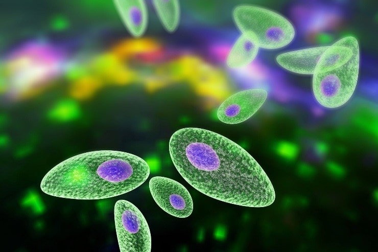
One of this month’s articles is focused on Toxoplasmosis, a protozoal infection that can cause clinical disease in domestic animals, and in particular to the diagnostic tests that can be requested at BattLab to diagnose the infection and monitor treatment.
Diagnosis of toxoplasmosis. The diagnosis of T. gondii infection or toxoplasmosis may be established by serologic tests, polymerase chain reaction (PCR) and cytological/histological demonstration of the parasite. Below you will find a schematic summary of all these tests with some more details that will be helpful in making the best choice.
1. Serologic testing
- IgM antibodies appear sooner after infection than IgG antibodies but generally do not persist past 3 months after infection. Therefore, increased IgM titers are consistent with recent infection. Persistent IgM titers (>4 months) have been documented in cats coinfected with FIV and in cats with ocular toxoplasmosis. Usually, IgM antibodies have a higher positive predictive value than IgG for clinical feline toxoplasmosis.
- IgG antibodies appear by the fourth week after infection and may remain increased for years during subclinical infection. To be useful, IgG titers must be measured in paired sera from the acute and convalescent stages (3–4 weeks apart) and must show at least a 4-fold increase in titer. Additionally, CSF and aqueous humor may be analysed for the presence of tachyzoites or anti-T gondii antibodies. By the time IgG antibodies are detected in feline sera, the oocyst shedding period has usually been completed.
Antibody test results alone cannot be used to make a diagnosis of toxoplasmosis. However, the following combination can be used to make a presumptive antemortem diagnosis:
- Demonstration of antibodies in serum, which suggests infection by T. gondii
- Demonstration of an IgM titer 1:64 or a 4-fold or greater increase in IgG titer, which suggests recent or active infection.
- Clinical signs of disease referable to toxoplasmosis
- Exclusion of other common causes of the clinical syndrome.
- Positive response to appropriate treatment.
2. PCR testing
The detection of T. gondii DNA by PCR is appealing, due to high sensitivity and specificity of this method. The presence of a parasitaemia is seldom detected, therefore, PCR of aqueous humor and CSF is preferred over blood PCR. A positive result detects the presence of the organism in infected animals but will not necessarily distinguish acute from chronic subclinical encysted infection. In the case of a positive PCR result, Toxoplasma is the probable cause of the clinical signs with appropriate clinical history, haematological, biochemical and/or cytological findings.
3. Cytology and histology
Post-mortem, tachyzoites may be seen in both cytological and histological samples However, T gondii is morphologically similar to other protozoan parasites and therefore it must be differentiated from Sarcocystis species and Neospora caninum by other diagnostic tests (serology, PCR).
All these tests are offered by BattLab therefore do not hesitate to contact us if you need more information or if you are considering to send us a sample.