
After the last two exhausting years, probably you are all fed up with Coronavirus… however, there is a serious disease affecting ferrets we must shed some light on.
In domestic ferrets (Mustela putorius furo), coronaviruses are responsible for two distinct clinical conditions, namely epizootic catarrhal enteritis (caused by ferret enteric coronavirus) and ferret systemic coronavirus (FRSCV)-associated disease.
FRSCV resembles the dry form of feline infectious peritonitis virus (FIPV)-induced disease, which is caused by an enteritis-inducing strain of feline coronavirus (FCoV) that is believed to mutate and develop the ability to replicate within macrophages. In 2006, the disease was extensively described for the first time (Martinez et al. 2006) and since then, in recent years, reports of this infection have become more frequent.
The infection mostly affects juvenile and young adult ferrets. The initial presentation is generally diarrhoea, often progressing to haemorrhagic form. Lethargy, anorexia, weight loss or inability to gain weight in young animals are also early signs. Enlarged abdominal masses (i.e. enlarged spleen and lymph nodes) often can be palpated. Less frequent findings include hindlimb paresis and CNS dysfunction. This condition is characterized by a progressive nature with an average survival time following diagnosis of just over two months.
Haematologic findings are nonspecific and include nonregenerative anaemia, hyperglobulinemia, hypoalbuminemia, and thrombocytopenia. Serum protein electrophoretogram shows a polyclonal hypergammaglobulinemia.
Cytological findings of splenic and lymph node aspirates include marked infiltration of macrophages and epithelioid macrophages, with a smaller component of neutrophils overlying the typical mixed lymphoid population expected from reactive lymphoid tissue.
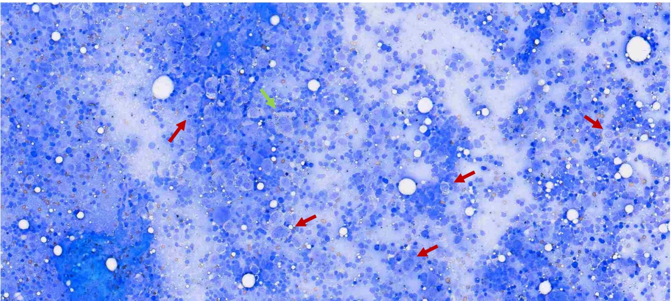 Picture 1: splenic aspirate from a ferret (Wright-Giemsa 50X). Red arrows indicate activated macrophages. The green arrow points towards an epithelioid macrophage.
Splenomegaly and hypergammaglobulinemia are commonly seen with Aleutian disease, lymphoma, multiple myeloma, chronic infection (e.g. Helicobacter sp), or chronic inflammation from inflammatory bowel disease (IBD).
Histologically, pyogranulomatous inflammation appears as a central area of necrotic cellular debris and degenerative neutrophils surrounded by epithelioid macrophages with occasional multinucleated giant cells and layers of lymphoplasmacytic infiltrates with a variable degree of fibrosis.
Definitive diagnosis of FRSCV-associated disease requires histopathology or PCR.
BattLab now offers the possibility to differentiate between enteric and systemic coronavirus, the latter considered proof of infection for FRSCV. Preferred samples include dry swab, faeces, EDTA blood or CSF. For more information, do not hesitate to contact us.