


Epidemiology & significance
Sporothrix spp. are saprophytic fungi that occur in their mycelial form in moist organic environments (e.g. soil, hay, water). They are opportunistic agents of mycoses. Four species within the Sporothrix schenckii complex are of pathogenic importance: S. schenckii, S. brasiliensis, S. globosa, and S. luriei. S. schenckii is distributed worldwide and is the most frequently detected species in Central Europe, whereas S. brasiliensis is endemic in some South American countries and represents a significant health risk for humans and animals there.
Disease in animals such as dogs, cats, horses, and in humans occurs most frequently in temperate and tropical climates. In Europe, prevalence is currently low and infections are generally caused by Sporothrix schenckii. Humans may become infected through wound contamination (skin injuries, e.g. via contaminated soil, thorns, wood splinters). In addition, infected cats (via scratches or bite wounds or direct contact with secretions) represent another important source of infection. Sporotrichosis is therefore considered a relevant zoonosis, and appropriate protective measures are recommended.
Clinical presentation of feline sporotrichosis
Feline sporotrichosis occurs most commonly in intact male free-roaming cats, following scratch or bite wounds from infected cats (asymptomatic carriers are possible). Typical sites include the head, limbs, and base of the tail (common targets during fights). In rare cases, infection may also occur via inhalation of the pathogen. Three clinical forms are recognised: cutaneous, mucocutaneous, and disseminated. The majority of cases are confined to the skin and subcutaneous adipose tissue, sometimes involving the lymph nodes. The mucocutaneous form is characterised by additional involvement of mucous membranes (nasal, conjunctival, oral, genital). In the less common disseminated form, systemic spread of the pathogen occurs, potentially affecting organs (particularly the lungs) and bones. In the cutaneous form, multifocal, raised, crusted, and alopecic plaques and nodules are observed, often ulcerated (Fig. 1). Lesions are frequently limited to the site of pathogen entry, as an intact host immune response prevents further dissemination. However, haematogenous spread (e.g. secondary to a primary respiratory form) or grooming behaviour may result in more extensive cutaneous dissemination.
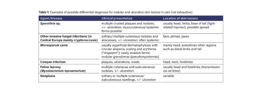
Although infections are rare in Europe, sporotrichosis should be considered, particularly in male free-roaming cats with ulcerative skin lesions, especially when a bacterial cause has initially been assumed and antibiotic therapy has been unsuccessful. Some possible differential diagnoses are summarised in Table 1.
As sporotrichosis is a zoonosis, handling of cats with ulcerative skin lesions should be carried out only with protective clothing and medical gloves, and precautions should be taken to prevent scratches and bite injuries. It should also be noted that some of the pathogens listed in Table 1 possess zoonotic potential.
For the clinical course and zoonotic potential, the complex interplay between virulence factors, or the pathogenicity of the organism (Sporothrix brasiliensis > Sporothrix schenckii), on the one hand, and predisposing host factors on the other, is decisive. Immunosuppression caused by retroviral infections or corticosteroid therapy may play a facilitating role. Other factors, such as regular deworming, may be associated with a stronger immune response and therefore milder disease and reduced zoonotic potential.
Disseminated forms are rare and may develop from cutaneous-lymphatic forms or following inhalation of the pathogen, depending on the factors mentioned above. Signs of systemic disease are often non-specific (fever, lethargy, anorexia) and, if the respiratory tract is involved, may include respiratory signs (sneezing, nasal discharge, dyspnoea). Respiratory signs may occur before the appearance of skin lesions or even in the absence of visible external changes.
Diagnostics
When sporotrichosis is included in the differential diagnosis of nodular or ulcerative skin lesions, the following diagnostic methods are useful:
Cytology
Cytology represents a relatively sensitive diagnostic method for the visualisation of Sporothrix. In smears from impression preparations of ulcerated skin lesions or fine-needle aspirates from nodules, the yeasts can be identified using routine cytological stains (e.g. Romanowsky-type stains) or special stains (such as the PAS reaction). Suspicious samples should be further examined by culture to confirm the diagnosis.
Histology
Histopathological examination is useful in non- ulcerated skin nodules. Histopathologically, feline sporotrichosis presents as diffuse or (multi-)nodular pyogranulomatous dermatitis and panniculitis, with variable degrees of ulceration and necrosis. The lesions typically contain numerous organisms. In tissue, Sporothrix spp. are present in yeast form, ranging from pleomorphic to round or cigar-shaped, and can be observed either freely within necrotic areas or within the cytoplasm of macrophages. With the aid of additional special stains (PAS reaction, Grocott stain), the yeasts can be visualised more clearly, particularly when only small numbers of organisms are present.

Culture
Culture is the diagnostic method of choice for confirming suspected cases. The laboratory should always be informed of the clinical suspicion in advance, as sporotrichosis is rare.
Suitable samples include exudates (preferably obtained from deeper areas of the skin lesions) or, preferably, tissue samples in order to avoid false- negative results. In disseminated cases, blood culture is recommended. In respiratory forms, additional suitable samples for culture include nasal swabs or bronchoalveolar lavage. The highest diagnostic accuracy is achieved through histological examination, including special stains, in combination with mycological culture. Specific detection methods such as PCR are offered by specialised laboratories.
Therapy & Prognosis
For the treatment of the cutaneous form of feline sporotrichosis, systemic therapy with itraconazole has proven effective, administered for at least two months or for one month beyond clinical resolution. A detailed treatment guideline can be found, for example, in the article by Rodrigues et al. mentioned below. Owner compliance is a key determinant of prognosis. With consistent treatment and in the absence of adverse effects from the medications used, the prognosis is good. In contrast,
disseminated infections or premature discontinuation of therapy are associated with an unfavourable prognosis.
Dr. Karen Lampe
Our diagnostic services for sporotrichosis include:
– Cytology
– Histopathology (including special stains)
– Mycology
Further literature:
Barrs VR, Beczkowski PM, Talbot JJ, Hobi S, Teoh SN, Muguiro DH, Shubitz LF, Sandy J. Invasive fungal infections and oomycoses in cats. 1. Diagnostic approach. J Fel Med Surg. 2024 Jan. 26 (1-22): doi: 10.1177/1098612X231219696

Gremião IDF, Miranda LHM, Reis EG, Rodrigues AM, Pereira SA. Zoonotic epidemic of sporotrichosis: cat to human transmission. PLoS pathogens, 2017 Jan. 13(1), doi:10.1371/1006077

Lloret A, Hartmann K, Pennisi MG, Ferrer L, Addie D, Belak S, Boucraut-Baralon C, Egberkink H, Frymus T, Gruffydd-Jones T, Hosie MJ, Lutz H, Marsilio F, Möstl K, Radford AD, Thiry E, Truyen U, Horzinek MC. Sporotrichosis in cats. ABCD guidelines on prevention and management. J Fel Med Surg. 2013 Jun. 15 (619-623): doi: 10.1177/1098612X13489225

Rodrigues EMF, Passos Cortez L, Passos Cortez L, Oliveira da Costa RC, Barbosa Costa S, Bezerra de Morais N, Ferreira TC. Advances in the therapeutic approaches to feline sporotrichosis: a review. Rev Bras Hig San Anim. 2025 Jun. 19 (1): 2. doi: 10.5935/1981-2965.20240042